Category Archives: Sickle cell complications

Leg Ulcers and SCD

Leg ulcers usually occurs on the lower part of the leg. They happen more often in males than in females and usually appear from 10 through 50 years of age. A combination of factors cause ulcer formation, including trauma, infection, inflammation, and interruption of the circulation in the smallest blood vessels of the leg.

Leg ulcers may be classified as acute or chronic according to their duration, however, there is no consensus as to a specific length of time to define chronicity. An acute ulcer usually should heal in less than a month. Among chronic ulcers, a duration of six months seems to define the most recalcitrant ulcers. It is not uncommon for ulcers to last many years, often closing and re-opening repeatedly times.

Leg ulcers are painful and often disabling complications of SCD. They tend to be indolent, intractable and heal slowly over months to years. The pain may be severe, excruciating, penetrating, sharp and stinging in nature. In most patients, oral or parenteral opioid analgesics are needed to achieve some pain relief. Leg ulcers are more common in males in some studies and their incidence increases with age.

Leg Ulcers tend to be difficult to treat successfully, healing slowly over months or years. They can severely disrupt quality of life, increase disability, require extended absence from the workplace, and place a high burden of care on healthcare systems.

Treatment
Leg ulcers can be treated with medicated creams and ointments. Leg ulcers can be painful, and patients can be given strong pain medicine. Management of leg ulcers could also include the use of cultured skin grafts. This treatment is provided in specialized centers. Bed rest and keeping the leg (or legs) raised to reduce swelling is helpful, although not always possible.
Treatment of leg ulcers includes wound care using wet to dry dressings. With regular and good localized treatment, many small ulcers may heal within a few months. Leg ulcers that persist beyond 6 months may require other modalities including, among other things, blood transfusion, skin grafting, Unna boots, zinc sulphate, hyperbaric oxygen, arginine butyrate, topical herbal applications, etc. Unfortunately success or failure of these measures has been anecdotal in nature without the advantage of controlled clinical trials to identify the best approach to management.
Principles of management of leg ulcers include education, protection, infection control, debridement and compression bandages. Debridement could be surgical, medical or biological. Osteomyelitis may complicate chronic leg ulcers, especially those with deep wounds, and it is advisable to rule out this complication with a bone scan or MRI and bone biopsy if needed. Rarely, a leg ulcer is the cause of systemic infections, and occasional deaths attributable to sepsis secondary to a leg ulcer infection have been reported. Also exceedingly rare is the need for amputation of the leg or the foot affected by the ulcer.
Nursing Considerations
Provide education to patient:
Avoid injury especially to feet, ankles and legs
To wear socks and appropriate fitting shoes
Use insect repellants
Avoid prolonged periods of time on feet
Do not allow needle sticks to lower extremities
Treat minor trauma promptly
Do not smoke
Teach patient treatment and protective measures
Types of dressings and changing techniques
Using aseptic technique when dressing wound
Elevate legs as much as possible
Eat a nutritious, well balance diet and take zinc supplements as ordered
Reinforce that compliance with treatment is the most important determinant of successful healing
Educate about judicious use of narcotics for this chronic pain and alternative pain control methods
Teach patient to avoid trauma, keep skin lubricated, leg as often as possible and to wear elastic support hose after the ulcer area heals to prevent recurrence
Provide counseling if patient is experiencing self/body image issues interfering with therapy
Prevention
Prevention of leg ulcers is very important. Patients must be instructed to treat minor trauma around the ankles promptly. Insect repellants and protection from chiggers is important. Edema should be treated with elevation of the legs or elastic stockings.
Once the ulcer heals, the patient should be instructed to avoid trauma to area, lubricate the area with skin moisturizers, and treat edema with leg elevation and elastic support. In our experience, edema is the single most common antecedent to recurrence of a leg ulcer.
Patient and Parent Education
Prevention of ulcers must be stressed beginning in childhood. The patient undergoing treatment must be given complete instructions and education about the normal course of treatment. The long-term nature of the ulcers and the importance of consistent and regular care must be stressed. Painful ulcers require education about judicious use of narcotic analgesics for chronic pain. Once the ulcer is healed all of the method of prevention must be reinforced with each visit.

wpid-wp-1445231628916.png

Priapism and Sickle cell

It is often said that sickle cell disease does not kill. This series will focus on breaking down and understanding each of the complications that might develop from sickle cell, so that you can know how to prevent this, catch it early and deal with it. Knowledge is power!
Priapism refers to a prolonged penile erection (e.g., lasting longer than 4 hours) unrelated to sexual stimulation or desire. There are 2 types of priapism: Low flow priapism [LFP]) results from reduced venous outflow from the erectile tissue (usually due to sickle cells clumping up the penile artery), and this is the most common type that affects sickle cell patients. High flow is usually unrelated to sickle cell, so I won’t bother going into it.
Risk Factors: 38-42% of men with sickle cell (or any variance eg thalassemias) will have LFP at least once during their lifetime. LFP usually peaks between the ages of 5-10 and 20-50. Use of recreational drugs like Cocaine, Marijuana and alcohol further increase your risk.
How do I know if I have Priapism?
If you have an erect, rigid, painful penis and no feelings of arousal, then you probably have priapism.
Besides the fact that priapism is uncomfortable, if left untreated, LFP can result in hypoxia (lack of oxygen to cells) and acidosis (increase of lactate acid in cells), tissue damage, and permanent erectile dysfunction (Eek!) and is considered a urologic emergency. So please, if you do have priapism, visit your doctor or go to the ER to get it treated! If you have priapism for 24 hours, you will become permanently impotent!
Treatment:
The first treatment option is to place ice packs to the penis and perineum to reduce swelling. With SCD patients, blood transfusions or exchange transfusions are given to increase the hematocrit value to greater than 30%. Hydration through IV fluids is given to thin out the blood. If this does not resolve LFP, the next step is an injection of Terbutaline, and if this is unsuccessful, another drug called Phenylephrine could be administered. In some cases, the doctor can draw out the blood from the penis with a needle, followed by a saline irrigation (water follows salt which is then excreted by the kidneys reducing the pressure). If these treatment strategies fail, you might have to have surgery to create a shunt that diverts the blood or a bypass of the blockage.
There are adverse effects related to the medications that will be injected. Report to your doctor if you have a headache, hypertension, dizziness, slow heart beating, fast heart rate, palpitations in the chest, signs of infection, bleeding from the site, or any changes to your penis.
How do I prevent Priapism?
There is very little you can do to prevent LFP. The best tips are to avoid triggers of your LFP (certain medications like anti-anxiety pills, Viagra, Levitra, and Cialis, anticoagulants, antihypertensives, antidepressants, antipsychotics, tranquilizers, antihistamines, cocaine, marijuana and alcohol). Drink at least 2 liters of water a day. Hydration works both ways to prevent crises and prevent cells from clumping together. For some reason, Glutamine and Arginine have caused some people to have LFP, even though in some it has the opposite affect. You might have to learn how to give yourself injections in the penis if this becomes a permanent recurrent problem.
Okay warriors, that is a brief synopsis of priapism. It is not something to be scared of, and many males with sickle cell have had it and will have it. So just follow these tips, put some ice on it, and if it doesn’t go away in a couple of hours, head to the ER/doc to get it taken care of.
Remember, knowledge is power. Be well.

ACS and Sickle cell

We all have heard about the dreaded Acute Chest Syndrome (ACS). Some of us have had it, and some sickle cell warriors have died from it. But what exactly is acute chest syndrome?
What is ACS? Acute chest syndrome (ACS) is a pulmonary illness defined by a new infiltrate on chest radiograph in combination with at least 1 clinical sign or symptom (chest pain, cough, wheezing, fast respiration rate, fever). Fever and cough are the most common presenting symptoms in children, while chest pain, shortness of breath, and chills are common in adults.
Many people state that it feels like you can’t get any air, as if you are suffocating, and struggling for every precious breath. It feels to some like an elephant sitting on your chest, and the pressure is compressing your lungs and making breathing an impossibility. Of all the complications, having your breath stolen from you, having every second a struggle for oxygen, this is by far the scariest, most panicky situation to ever be in.
Recurrent episodes are thought to contribute to chronic lung disease, pulmonary hypertension, and cor pulmonale.
Physiology: During a sickle cell crises, the sickled cells have problems passing through the tiny vasculature of the lung tissue. This causes a blockage which leads to injury which then starts the inflammation, hypoxia, and lung injury cycle. This cycle continues on and on similar to what goes on in Acute Respiratory Distress Syndrome until large parts of the lung is injured and unable to process oxygen. Eventually if this process continues, the patient will be completely unable to breathe on their own and may even need to be intubated and mechanically ventilated (which is what happened to me).
What causes Acute Chest Syndrome? ACS is associated with all genotypes of sickle cell but occurs most frequently in patients with homozygous (SS) disease. The main causes of acute chest syndrome are pulmonary infections (like pneumonia, bronchitis, RSV, CMV, flu etc.), emboli (like fat clots or blood clots), and rib infarctions which is caused by splinting or rib breakage during a vaso-occlusive crises.
The other possible causes is what is called iatrogenic. Excessive narcotic use and excessive hydration have been proposed as 2 possible iatrogenic causes of ACS; however, both have limited supporting evidence. It has been suggested that atelectasis secondary to hypoventilation and poor respiratory effort with narcotic use might lead to ACS. In addition, it has been suggested that patients admitted for sickle cell pain crisis may develop pulmonary edema and subsequently ACS if hydration strategies are too aggressive. This is why alot of physicians err on the side of caution when prescribing fluids and narcotics to sickle cell patients.
How do you treat Acute Chest Syndrome? Treatment involves supportive care, incentive spirometry, broad-spectrum antibiotics for the infection, bronchodilators (to expand the airway and improve oxygenation), and early blood transfusion when indicated.
In short: Acute Chest Syndrome is a medical emergency, a serious complication that can be life-threatening. 4.6% of patients with ACS die from respiratory complications, and ACS can increase your length of stay by 5 days. Please, please my dear warriors! Prevent this from happening to you as much as possible. Use your incentive spirometer, because this helps to improve oxygenation, improve blood flow to lung tissue and expand the alveoli. The incentive spirometer has to be your best friend when you are admitted, regardless of whether you are in pain or not. The recommendation is 10 breaths every hour when awake, between the hours of 8am to 10pm. I have a habit that whenever I press my PCA button, I use the incentive spirometer.
If you are having any problems breathing during your sickle cell crises, be sure to mention the possibility of acute chest syndrome to your doctor, nurse or advocate to bring it to their attention. Early identification and aggressive treatment has given better outcomes and most people with ACS recover fully.

AVN and Sickle Cell

This series will focus on breaking down and understanding each of the complications that might develop from sickle cell, so that you can know how to prevent it, and how to catch it early.
What is Avascular Necrosis?

image

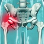
Avascular Necrosis (AVN), government name osteonecrosis, is defined as death of the bone due to reduced or limited blood supply to the bone tissue. The necrosis (or death), leads to a thick scar tissue developing, which then prohibits any further healing of the bone, leading to joint pain and loss of function.
The joints most often affected by AVN (in decreasing order of incidence) are the hip, knee, shoulder, ankle/foot (talus), wrist and elbow. This is because these sites are more prone to have less collateral (smaller veins) that can provide circulation to the affected area, and usually only have a single large blood vessel that delivers circulation. Sickled cells find it quite easy to clump up this one blood vessel, and the risk for AVN is developed.
Risk Factors: AVN occurs in individuals of all ethnicities; although those with sickle cell diseases are more prone to this complication. It is more common in men than women (ratio of 8:1), and develops most often in middle age. With SCD, patients develop AVN from age 12, and once you have developed it, there is an increased chance for getting AVN in the future. Alcohol use puts you at risk for AVN. Also, patients who are on dialysis, have lupus, or who routinely use steroids (like in the treatment of acute chest syndrome, and pulmonary hypertension, are at an increased risk of getting AVN). More than 50% of patients diagnosed with AVN require surgery 3 years after.
What Causes AVN in Sickle Cell? : When in crises, sickled cells float around clumping up in various parts of the body. This leads to tissue starvation and pain. The bone is a living structure, and requires blood circulation to provide oxygen and energy to all the cells. The arteries and veins are responsible for providing this nutrition. In the bone structure, there is usually only one vein and one artery that delivers blood to a certain area, and once this is clogged up by sickled cells, there are no alternate routes. This leads to death of the bone. Osteoblasts and osteocytes come in to curtail the spread of infection, and a thick scar tissue is developed on the blackened, dead area. This means that no healing or repair can be done once the sickle cells leave and circulation is restored. As a result, that part of affected bone is PERMANENTLY dead, and is unable to function normally.
How do you know if you have AVN? The main sign of AVN is pain in the bone that is there regardless of whether or not you are in crises. Usually this pain is in the joint between the hip and thigh bone, and can be felt when walking. There might be tenderness in other surrounding areas. You might find yourself unable to have a normal range of motion in the affected side. You might find yourself unable to bend over and tie your shoes. In some people, AVN is so advanced that you might have a slight limp or clicking in movement. In addition, if the bone compresses the nerve, there might be some numbness, tingling or lack of sensation in the affected extremity.
AVN of the hip sometimes shows up on an X-ray as a blackened area. However, unless your doctor suspects AVN he won’t routinely order this X-ray for you. You have to ask for it. In other parts of the body, the AVN might be so microscopic that it won’t show up on an X-Ray. CT scans or Bone scans are also used, but are not sensitive enough tests. This is why it’s important to ask for an MRI of the affected area. Although MRIs are expensive and there is an increased risk of radiation, they are the most effective with diagnosing AVN.
How do you treat it? There are 7 stages of AVN and the treatment plan is based on what stage of AVN you are in, whether it is symptomatic, and the severity of symptoms.
Joint/Hip Replacement: Since AVN affects the joints or hip, the dead section is completely removed and a metallic joint is placed in that works just like the dead bone should. Doctors tend not to be aggressive in suggesting Joint Replacement until AVN is in its advanced stages. This is because once you have a hip or joint replacement, you have to have another surgery every 10 years to replace the hardware. And you know every time you go under the knife, there is a risk of complications like infection and hardware rejection.
Core Decompression: The orthopedic surgeon will drill a hole into the affected bone that takes a chunk of the dead necrotic area out. This leads to less pressure on the bone. The thought is that this will then allow the bone to regenerate on its own, since the scar tissue has been removed and blood circulation has been restored. This often works in intermediate stages, promotes healing and provides relief from the pain. It is 35-95% successful in treating AVN patients, especially those in pre-collapse.
Osteotomy: This procedure involves rotating one of the joints, so that the joint is no longer resting on a dead, painful area. It is not as effective as core decompression, and as a result is rarely suggested.
Bone Graft: This involves melding a new piece of bone gleaned from a healthier part of the body, and grafting it unto the dead area, thereby promoting circulation and healing. Bone graft is often done with core decompression and in small areas, is about 70-91% effective in mild to moderate AVN. This option is great because there is a possibility for complete bone healing, especially in the femoral head. In addition, there is a reduced risk of infection from hardware placed during joint replacement. However, the recovery time is longer than with joint replacement and there is less complete pain relief.
Pain Management: In mild AVN, the conservative treatment is pain management through NSAIDs like Motrin, Aleve or other pain-killers and analgesics. If the pain is under control, then you can engage in flexibility and joint exercises which might help to promote circulation to the bone.
Alternative Remedies: To prevent and treat AVN at home, it is recommended to:
Engage in activities and increase circulation in the bone like low impact exercises, walking, yoga, swimming
Take supplements that promote bone growth like calcium, vitamin D, and carao
REFERENCE: TOFFIERI, J.K. & GILLILAND, W. AVASCULAR NECROSIS: EMEDICINE RHEUMATOLOGY. PUBLISHED DECEMBER 19, 2009. HTTP://EMEDICINE.MEDSCAPE.COM/ARTICLE/333364-OVERVIEW